


Forskere fremskynder billeddannelsesteknikker til at fange strukturer af små molekyler
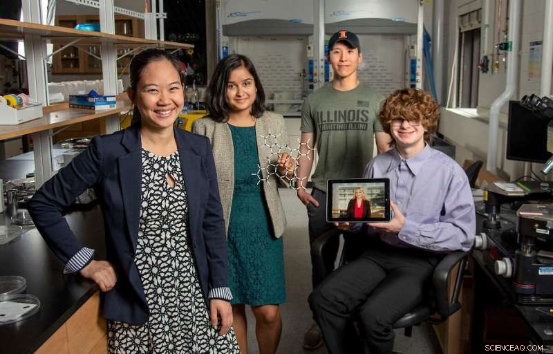
Pinshane Huang, yderst til venstre, slutter sig til, Priti Kharel, næst fra venstre, Justin Bae og Patrick Carmichael, yderst til højre, på Materials Research Laboratory. Billedet på tabletten er Blanka Janicek. Kredit:Heather Coit/The Grainger College of Engineering
En forskningsindsats fra University of Illinois Urbana-Champaign ledet af Pinshane Huang accelererer billeddannelsesteknikker for at visualisere strukturer af små molekyler klart - en proces, som engang troede umulig. Deres opdagelse frigør uendeligt potentiale i at forbedre hverdagens anvendelser - fra plastik til lægemidler.
Lektor ved Institut for Materialevidenskab og Teknik er gået sammen med hovedforfatterne Blanka Janicek, en '21-alumne og post-doc ved Lawrence Berkeley National Laboratory i Berkeley, Californien, og Priti Kharel, en kandidatstuderende ved Institut for Kemi, at bevise den metodologi, der gør det muligt for forskere at visualisere små molekylære strukturer og accelerere nuværende billeddannelsesteknikker.
Yderligere medforfattere inkluderer kandidatstuderende Sang hyun Bae og studerende Patrick Carmichael og Amanda Loutris. Deres peer-reviewed forskning er for nylig blevet offentliggjort i Nano Letters .
Holdets indsats afslører molekylets atomare struktur, så forskerne kan forstå, hvordan det reagerer, lære dets kemiske processer og se, hvordan man syntetiserer dets kemiske forbindelser.
"Strukturen af et molekyle er så fundamental for dets funktion," sagde Huang. "Det, vi har gjort i vores arbejde, er at gøre det muligt at se den struktur direkte."
Evnen til at se et lille molekyles struktur er afgørende. Kharel fortæller, hvor vigtigt det er, ved at give eksemplet med en medicin kendt som thalidomid.
Opdaget i 60'erne blev thalidomid ordineret til gravide kvinder til behandling af morgenkvalme og blev senere fundet at forårsage alvorlige fødselsdefekter eller i nogle tilfælde endda død.
Hvad gik galt? Lægemidlet havde blandede molekylære strukturer, den ene ansvarlig for behandling af morgenkvalme og den anden forårsagede desværre ødelæggende, negative virkninger for fosteret.
Behovet for proaktiv, ikke reaktiv videnskab har opfordret Huang og hendes elever til at forfølge denne forskningsindsats, der oprindeligt begyndte med ren og skær nysgerrighed.
"Det er så afgørende at præcist bestemme strukturerne af disse molekyler," sagde Kharel.
Typisk bestemmes molekylære strukturer med indirekte teknikker, en tidskrævende og vanskelig tilgang, der bruger kernemagnetisk resonans eller røntgendiffraktion. Endnu værre, indirekte metoder kan producere forkerte strukturer, der giver forskere den forkerte forståelse af et molekyles sammensætning i årtier. The ambiguity surrounding small molecules' structures could be eliminated by using direct imaging methods.
In the last decade, Huang has seen significant advancements in cryogenic electron microscopy technology, where biologists freeze the large molecules to capture high-quality images of their structures.
"The question that I had was:What's keeping them from doing that same thing for small molecules?" Huang said. "If we could do that, you might be able to solve the structure (and) figure out how to synthesize a natural compound that a plant or animal makes. This could turn out to be really important, like a great disease-fighter," Huang said.
The challenge is that small molecules are often 100 or even 1,000 times smaller than large molecules, making their structures difficult to detect.
Determined, Huang's students began using existing large molecule methodology as a starting point for developing imaging techniques to make the small molecules' structures appear.
Unlike large molecules, the imaging signals from small molecules are easily overwhelmed by their surroundings. Instead of using ice, which typically serves as a layer of protection from the harsh environment of the electron microscope, the team devised another plan for keeping the small molecules' structures intact.
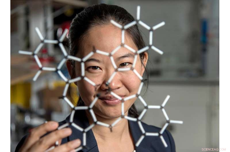
Pinshane Huang and her researchers discovered how to view structures in molecules, which opens a whole new realm of scientific possibilities. Credit:Heather Coit/The Grainger College of Engineering
How can you temper a molecule's environment? By using graphene.
Graphene, a single layer of carbon atoms that form a tight, hexagon-shaped honeycomb lattice, dissipates damaging reactions during imaging.
Stabilizing the small molecule's environment was only one issue the Illinois researchers had to manage. The team also had to limit its use of electrons, as low as one-millionth the number of electrons normally used, to illuminate the molecules.
Low doses of electrons ensure that the molecules are still moving enough for the researchers to capture an image.
"The way I like to think about it is the molecule doesn't like to be bombarded by higher-energy electrons, but we need to do that to be able to see the structure, and graphene helps dissipate some of that charge away from the molecule so that we can actually get a nice image of it," Janicek said.
Unfortunately, once captured, the molecules were nearly invisible in the image.
"When they take a low-dose image, it initially looks like noise or TV static—almost like nothing is there," Huang said.
The trick was to isolate the atomic structures from that noise by using a Fourier transform—a mathematical function that breaks down the small molecule's image—to see its spatial frequency.
"We took images of hundreds of thousands of molecules and added them together to build a single, clear image," Kharel said.
This averaging approach allowed the team to create crisp images of the molecules' atoms without damaging the integrity of any individual molecule.
"Month after month, week after week, our resolution improved," Huang said. "And then one day, my students came in and showed me the individual carbon atoms—that's a major achievement. And of course, it comes after all this deep knowledge that they have gained to design an imaging experiment and how to unlock data from what looks like nothing."
This collective discovery is paving the way for many more structural molecule imaging findings.
"There's been this whole field of small molecules that have been left out in the cold, so to speak. We're shining a light on how do we get there as a field? How do we make this thing that for us right now is so hard?" Huang said. "One day it won't be—that's the hope."
The Illinois researchers' efforts are the first big step in turning that dream into reality.
"One day, this will be the way we solve the structure of a small molecule," Huang said. "People will simply throw the molecule in the electron microscope, take a picture and be done."
That dream inspires Huang and her Illinois team to keep the course.
"That's potentially life-changing, and we've made it exist," Huang said. "We haven't yet made it easy, but imaging techniques like this will change so much of science and technology." + Udforsk yderligere
ESR-STM på enkelte molekyler og molekylebaserede strukturer
 Varme artikler
-
 Almindelige nanopartikler fundet at være meget giftige for det arktiske økosystemQueens University-forskerne Niraj Kumar og Virginia Walker ved siden af det udstyr, de brugte til at måle respirationen af mikrobesamfund, der lever i arktiske jordprøver. Måling af denne respirat
 -
 Forskere bruger polystyren til at gøre næste generations solpaneler endnu billigereKredit:University of Manchester Forskere fra University of Manchester bruger polystyrenpartikler frem for dyre polymerer til at lave den næste generation af solceller, som bruges til at lave solpa
 -
 Forskere opretter verdens mindste snemand (m/ video)Verdens mindste snemand måler 0,01 mm på tværs, med en næse på kun 0,001 mm. Kredit:National Physical Laboratory. (PhysOrg.com) - David Cox, en videnskabsmand i Quantum Detection -gruppen ved Nati
 -
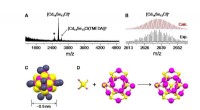 Strukturen af den mindste halvleder belystSammensætning og strukturel karakterisering af Cd14Se13-klyngen. A., B. Massespektre med høj opløsning. C. Overordnet molekylær struktur. D. Dannelse af Se-Cd14Se12 kerne-burstrukturen. E. Chlorider s
- Konvoj, appen Uber for Trucking, scorer $ 400 millioner i ny finansieringsrunde
- Højhastighedsopladningsenhed - succes i højkapacitets grafenbaserede superkondensatorer
- Efterligner enzymer, kemikere producerer store, nyttige carbonringe
- Oscar Madisons tilgang til solceller kan overstråle Felix Ungers design
- Har kometen indvirket på at starte livet på Jorden?
- Billede:Orion-rumfartøjet skrider frem med installation af modul til at teste fremdriftssystemer